Raman Spectroscopy: a technique to detect bacteria
Author: Bogdan Parakhonskiy, from Ghent University
What is Raman Spectroscopy?
It is a non-destructive chemical analysis technique which uses the interaction of light with matter to obtain information about the composition or characteristics of a material.
Raman spectroscopy exploits the effect of laser light scattering upon interacting with a sample. During this process, energy is dissipated between incident photons and the sample molecules. Energy scattered during this process corresponds to specific molecule vibrations. The fact that Raman scattering components can be linked with the molecular composition of the investigated sample in a label-free manner – makes this spectroscopic technique highly attractive for various analytes.
Following the improvement of the method to surface-enhanced Raman spectroscopy (SERS) is a powerful analytical method since it enables several orders of magnitude enhancement of Raman signal. Enhancement of the signal is generated by plasmonic nanoparticles that are in most cases made up of gold or silver. The localized surface plasmon resonance of electrons occurs upon light illumination on the surface of such metal moieties within plasmonic nanoparticles.
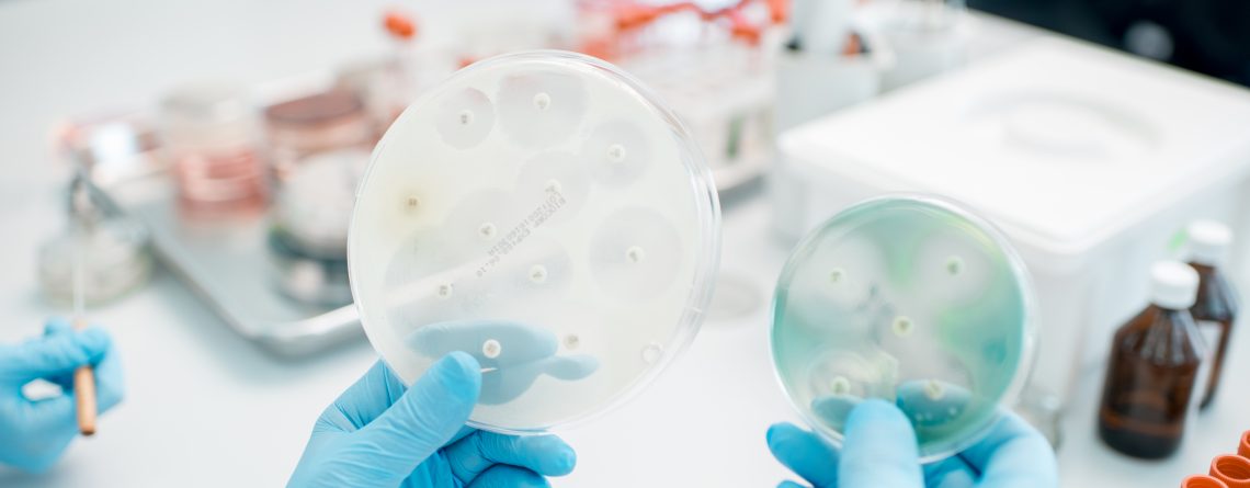
Raman spectroscopy for characterization applicable to bacteria can be used for the following applications: 1) determination of bacteria species; 2) determination of dead-alive states of bacteria; 3) determination of the bacterial metabolic activity, and even 4) distinction between different phenotypes of bacteria.
The most significant advantage of Raman spectroscopy is its label-free nature of detecting molecules and, therefore, bacteria and biofilms. The applicability of this approach in a native environment (for example, an aqueous solution) makes this technique particularly attractive for biological applications. In addition, it is a reproducible technique, which can also be implemented in a portable device, where both Raman scattering (easily applicable in aqueous samples) or infrared absorption (where OH vibrations should be taken into consideration) of vibrational bonds can be probed.
References:
[1] E. Lengert, M. Saveleva, A.A. Abalymov, V. Atkin, P.C. Wuytens, R. Kamyshinsky, A.L. Vasiliev, D.A. Gorin, G.B. Sukhorukov, A.G. Skirtach, B. V. Parakhonskiy, Silver Alginate Hydrogel Micro- and Nanocontainers for Theranostics: Synthesis, Encapsulation, Remote Release, and Detection, ACS Appl. Mater. Interfaces. 9 (2017) 21949–21958. https://doi.org/10.1021/acsami.7b08147.
[2] E. Lengert, B. V. Parakhonskiy, D. Khalenkow, A. Zečić, M. Vangheel, J.M. Monje Moreno, B.P. Braeckman, A.G. Skirtach, Laser-induced remote release in vivo in C. elegans from novel silver nanoparticles-alginate hydrogel shells, Nanoscale. 10 (2018) 17249–17256. https://doi.org/10.1039/C8NR00893K.
[3] E. Lengert, A.M. Yashchenok, V. Atkin, A. Lapanje, D.A. Gorin, G.B. Sukhorukov, B. V. Parakhonskiy, Hollow silver alginate microspheres for drug delivery and surface enhanced Raman scattering detection, RSC Adv. 6 (2016) 20447–20452. https://doi.org/10.1039/C6RA02019D.


Comments (3)
Everything is very open with a very clear description of the challenges. It was really informative. Your site is very helpful. Many thanks for sharing!
I am not certain the place you’re getting your information,
but great topic. I needs to spend a while studying more or figuring out more.
Thank you for excellent info I was searching for this information for my
mission. ronaldo drakt barn
JeanaScha psg trøje|juventus trøje SterlingT
Excellent goods from you, man. I’ve take note your stuff prior to and you are just extremely excellent.
I actually like what you’ve got right here, certainly like what you’re saying
and the best way through which you assert it. You make it enjoyable and you continue to take
care of to keep it sensible. I can’t wait to learn far more from you.
That is really a wonderful site.